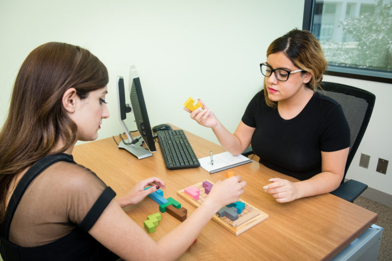
Based on the latest scientific findings in the field of neuroscience, Dr. Fotuhi has developed tailored programs for recovery from concussion and memory loss. One aspect of those plans involves brain coaching, a one-on-one session with a dedicated, trained advocate. During sessions, patients have the opportunity to speak with their brain coach about attainable solutions to enhancing memory, reducing stress, improving organizational skills, and discovering creative ways to overcome challenging life circumstances.
Based on his 25 years of research, clinical work, and teaching in neuroscience at Johns Hopkins and Harvard Medical School, Dr. Majid Fotuhi has summarized this literature in his scientific peer-reviewed articles (and books) and formulated a “Brain Fitness Program” to use effective behavioral modification techniques for enhancing cognitive function. This program produces remarkable results in patients with memory loss, post-concussive syndrome, and mild cognitive impairment.
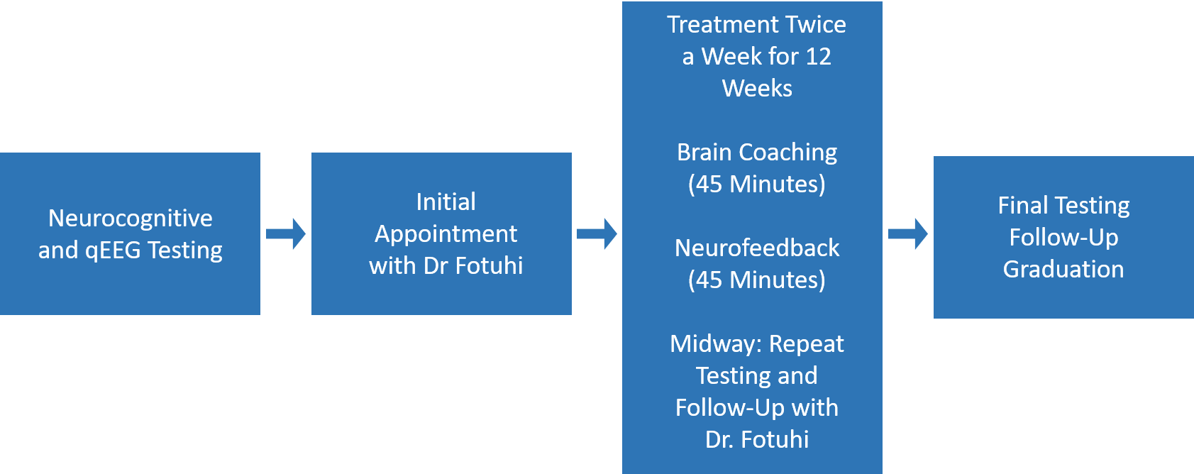
The Brain Fitness Program consists of a comprehensive brain health assessment followed by a series of interventions designed to optimize memory and overall cognitive performance. Over a period of 12 weeks, patients first undergo a detailed neurological evaluation to determine all of the potential causes for cognitive deficits and brain atrophy. They then start an intensive set of treatment sessions that enhance cognitive function, reduce anxiety, improve sleep, and focus on lifestyle factors that are shown to be critical for prevention of late-life dementia.
The program has eight important interventions, starting with patient’s sense of “purpose in life” and their desire for peak brain performance in order to achieve a personal goal:
Published in NeuroRegulation (2017), about the benefits of neurofeedback for treating anxiety and depression
Published in the Journal of Prevention of Alzheimer’s Disease (2016) , about our 12-week Brain Fitness Program
Published in Nature Reviews Neurology (2012), about how to grow the memory part of our brain
Published in Nature Reviews Neurology (2009) , about how late-life memory loss has many different treatable etiologies
Learn more the scientific basis for the different components of the brain coaching in our Brain Fitness Program.

The manuscript with results from patients who completed the Brain Fitness Program was presented at the Alzheimer’s Association International Conference in Washington DC, on July 18th, 2015. Below are the figures from this manuscript. They show that 84% of patients experienced high-impact improvements in their cognitive function, 97% had high-impact or moderate-impact improvements in their brain wave normalization. Among those who had before-and-after brain MRIs, 65% had remarkable reversal of atrophy (12%) or actual growth (53%) in the size of their hippocampus.

Percent of patients in the Brain Fitness Program (n=127) who had statistically significant improvements in their cognitive function.
More information can be found in our 2016 study, published in the Journal of Prevention of Alzheimer’s Disease (2016) , about our 12-week Brain Fitness Program
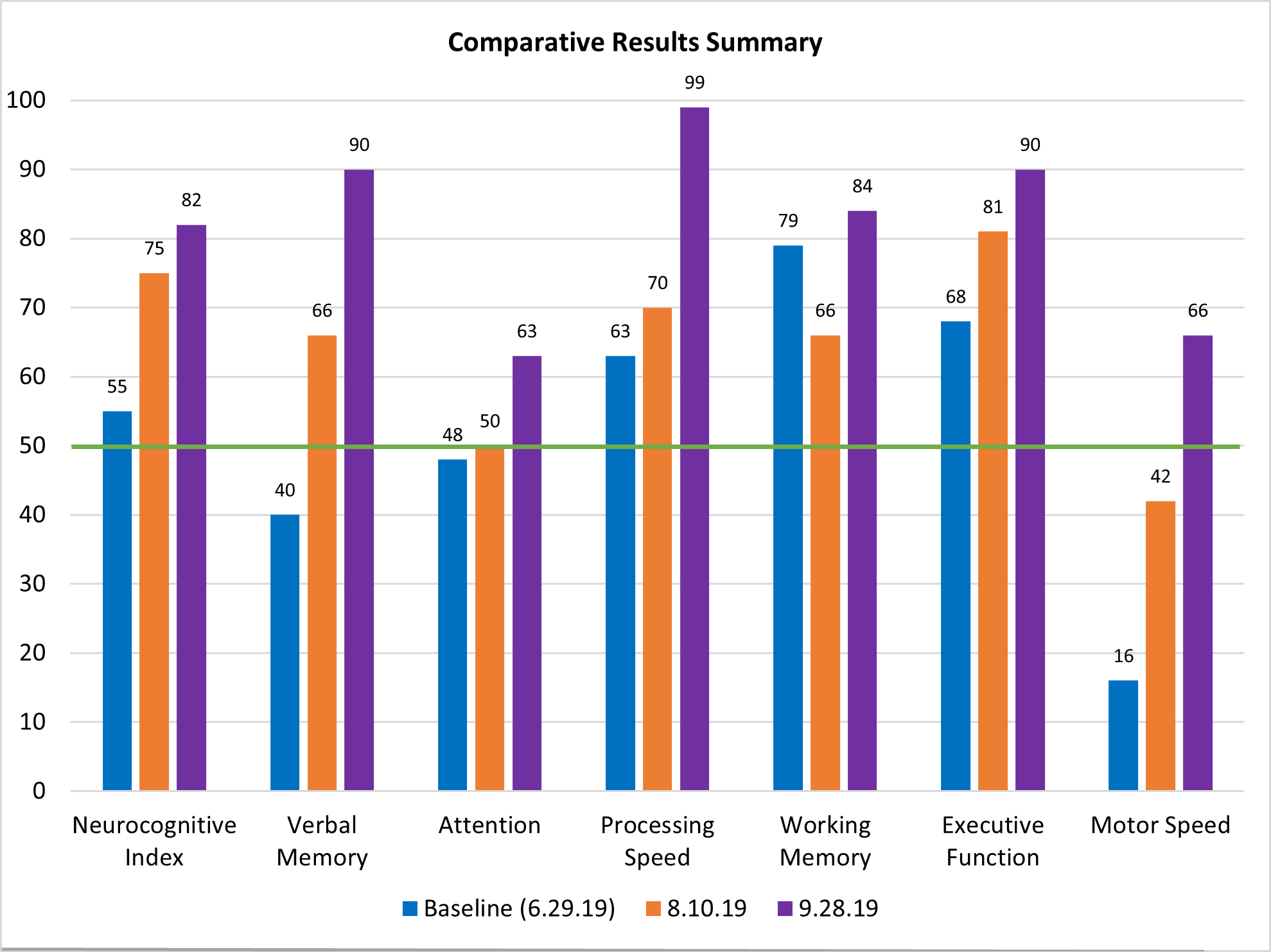
The cognitive results in one of the patients who completed the Brain Fitness Program shows remarkable improvements in her executive function, memory, and processing speed.
Patient’s baseline cognitive function is shown as percentile compared to others of her age and gender.
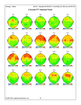
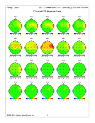
Brain mapping (Q-EEG) results in one of the patients who completed the Brain Fitness Program shows significant reduction (reduced red areas) in abnormally slow (theta) brain wave activity.
Q-EEG detects changes in a patient’s brain activity and compares it with a normative set of data. Statistically significant higher than normal or lower than normal EEG activities are shown in red or blue colors, respectively. Post-program Q-EEG (B) in this patient showed significant improvement toward normal (46%) from baseline (A), through a reduction in excessive levels of slow theta waves in her right temporal lobe.
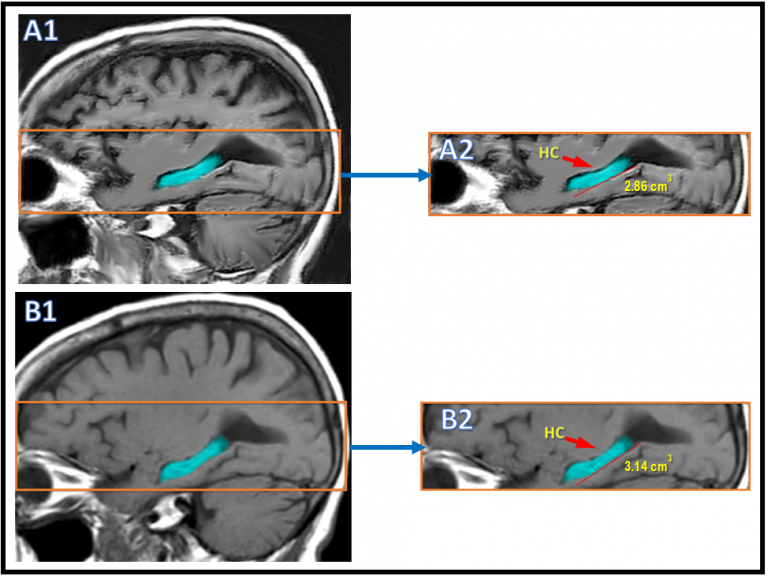
MRI results in one of the patients who completed the Brain Fitness Program shows significant enlargement in the size of hippocampus.
Post-program MRI with NeuroQuant showed an 8.6% growth in the volume of the hippocampus (HC). (A1): Baseline Brain MRI. (B1): Post-program Brain MRI. (A2): Higher magnification of baseline hippocampal volume. (B2): Higher magnification of the post-program hippocampal volume.
Get weekly science-based brain health tips delivered to your inbox.
© 2026 Dr. Majid Fotuhi | Legal